Mesenchyme Cells in the Sand Dollar Dendraster
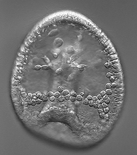
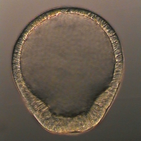
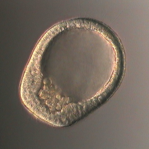
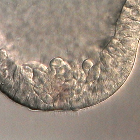
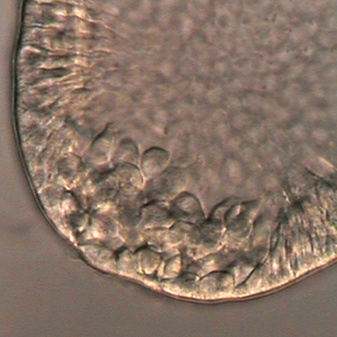
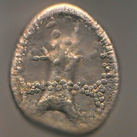
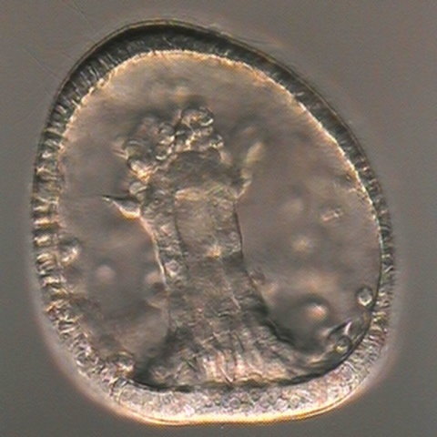
Echinoid embryos have two populations of mesenchymal cells with distinct fates and origins. Just before gastrulation the vegetal end of the blatula thickens and flattens (top left image). Some of the vegetal plate cells delaminate and accumulate in a heap just inside the blastocoel cavity (second through fourth images). These primary mesenchyme cells arrange themselve is a loose circlet around the archenteron as it invaginates, then fuse with each other and secrete the larval skeletal spicules. The very first triradiate spicules are faintly visible in the fifth image. Meanwhile, about halfway through gastrulation, secondary mesenchyme cells begin to emerge from the archenteron tip. Many of these cells will form coelomic pouches and some will form larval muscles.
|
|
|
|
|
|
Except for the embryo in the middle of the top row, these embryos were slightly flattened with a coverslip, on the one hand to stop the little buggers from swimming off before their pictures were taken and on the other hand to make it easier to capture the cells inside the blastocoel.