Cleaving Blastomeres in the Sand Dollar Dendraster
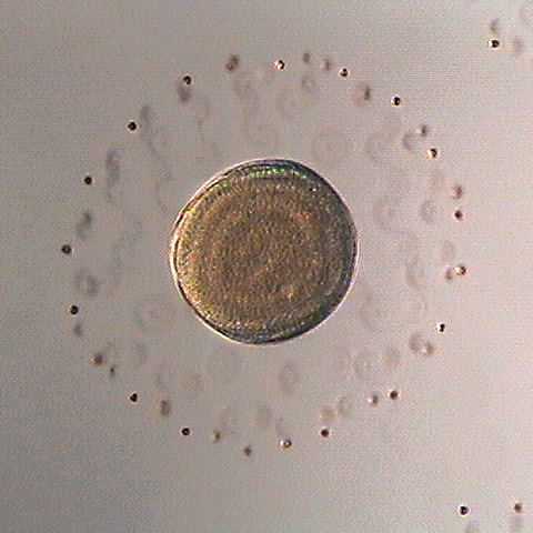
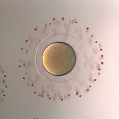
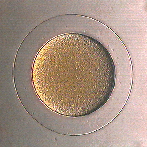
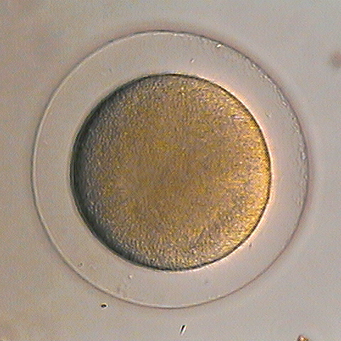
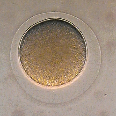
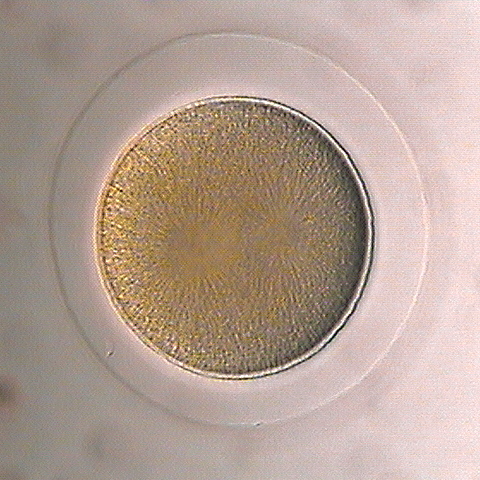
Dendraster eggs come out looking rather deflated and sad but surrounded by a beautiful corona of follicle cells embedded in the periphery of the jelly coat (left panel, below). Upon fertilization they round up quite nicely (middle panel). They have a fairly high fertilization envelope and a thin hyaline layer. Some minutes after fertilization it's usually possible to spot the sperm pronucleus (right panel, in the upper left of the cell).
|
|
|
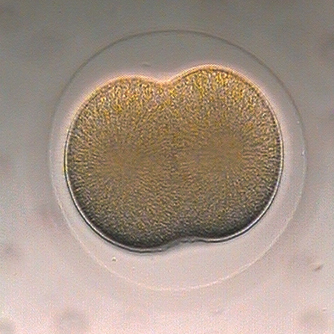
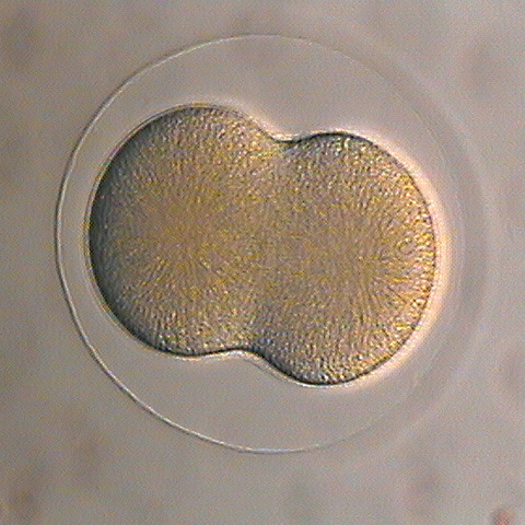
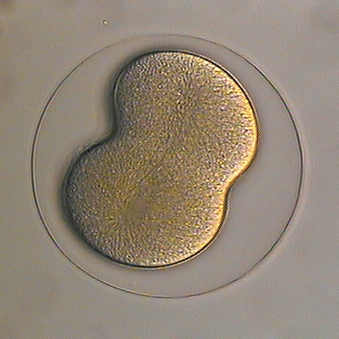
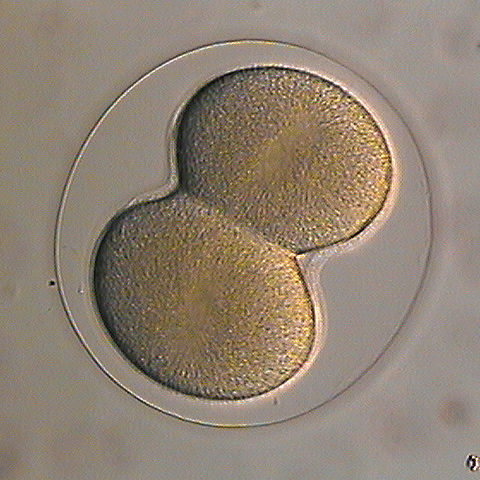
As the zygote approaches first cleavage the metaphase spindle asters become faintly visible (left-most panel, top row, below), and grow more prominent in anaphase (middle and right panel). As furrowing proceeds the asters become less and less clear, and nuclei reform near the center of each daughter cell (bottom row, middle panel).
|
|
|
|
|

|
|
|
|
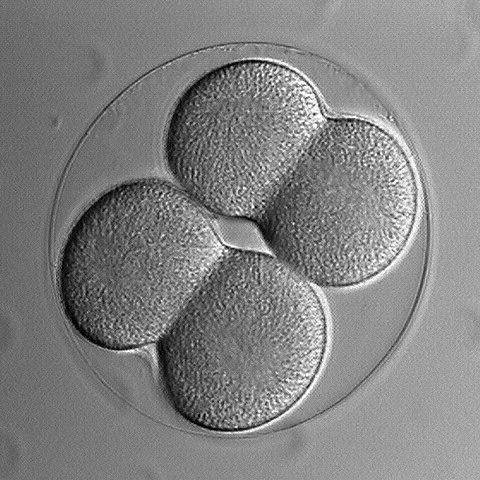
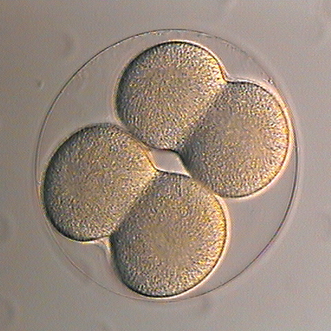
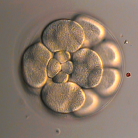
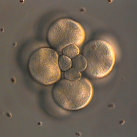
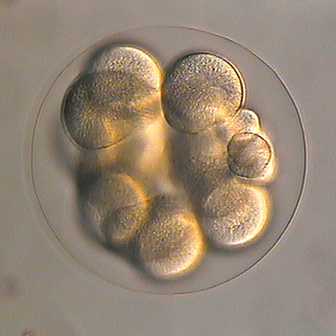
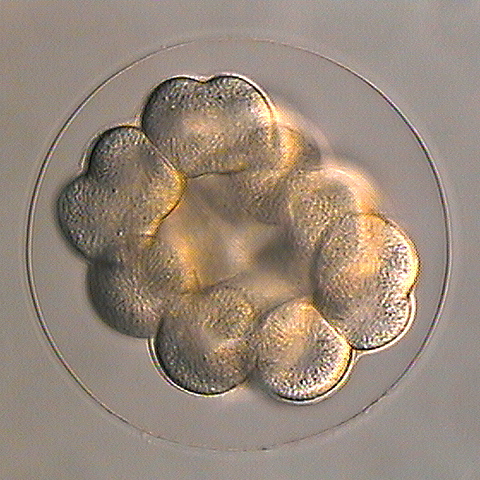
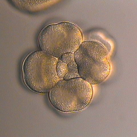
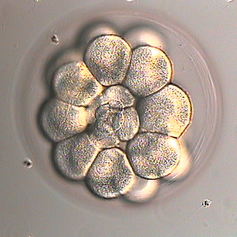
The first three divisions are equal, but at fourth cleavage the vegetal tier divides unequally, yielding four small micromeres and four large macromeres. The animal tier cells continue to divide equally. The macromeres divide equally at fifth cleavage (middle panel, bottom row, below) but the micromeres undergo another unequal division (left panel, bottom row).
|
|
|
|
|
|
Dendraster, like most echinoids, spawn when injected with isotonic KCl. A ripe female can make as much as 5 ml of eggs. They can be kept ripe in the lab throughout the summer. The embryos tolerate much warmer culture temperatures (up to 23°C) than a lot of other local species, and even tolerate an uncooled microscope stage as long as the lights aren't too high. Another nicety is that the fertilization envelope remains soft for up to a half hour after fertilization, and one can strip it off just by passing the eggs through 130 µm nitex mesh.