Embryogenesis in the Sea Squirt Boltenia
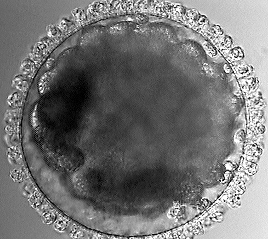
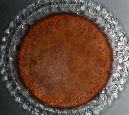
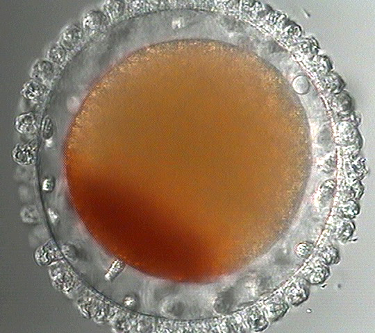
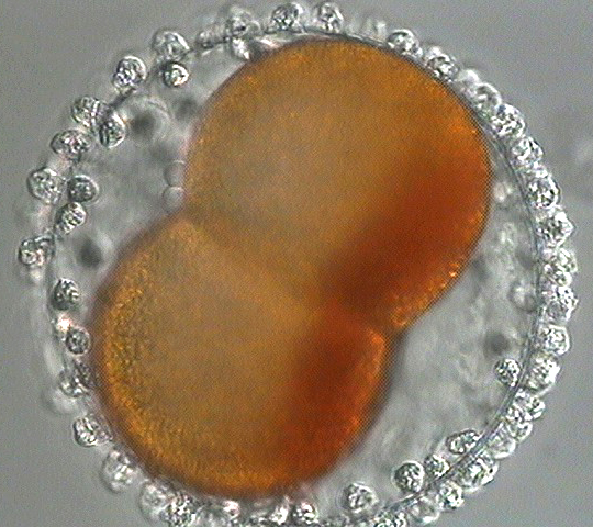
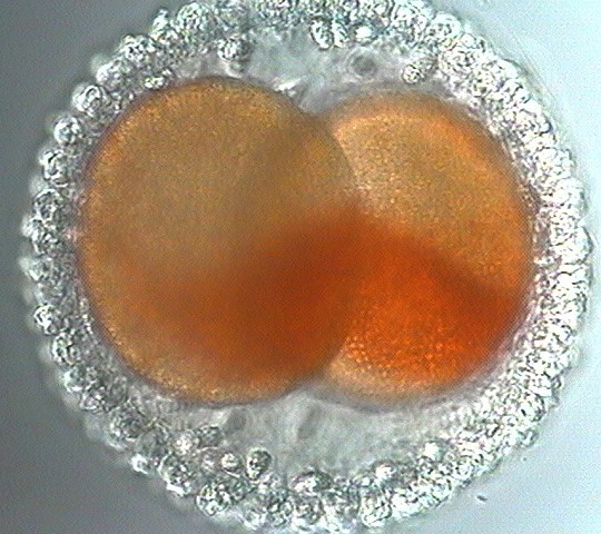
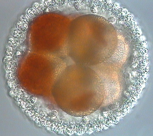
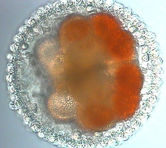
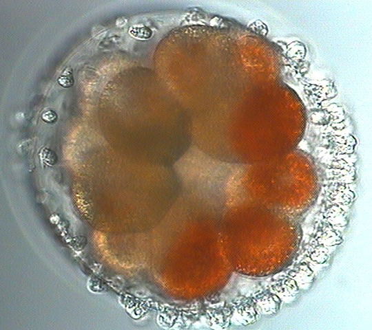
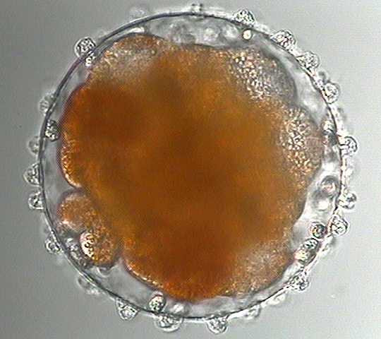
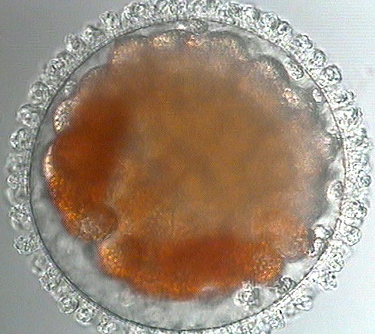
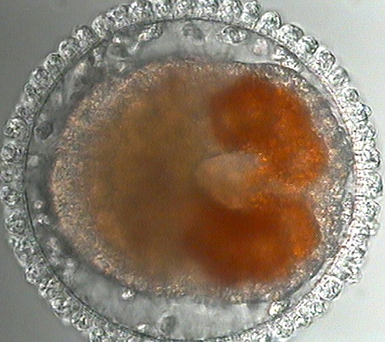
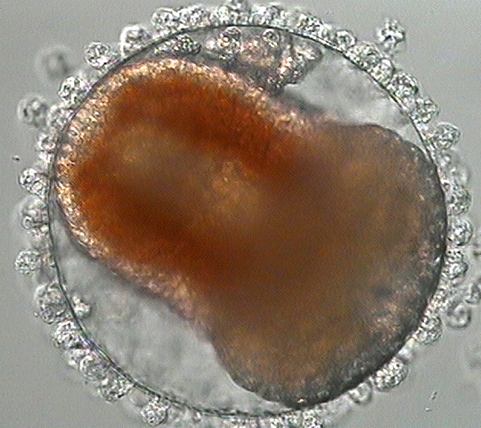
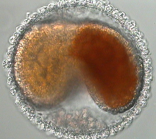
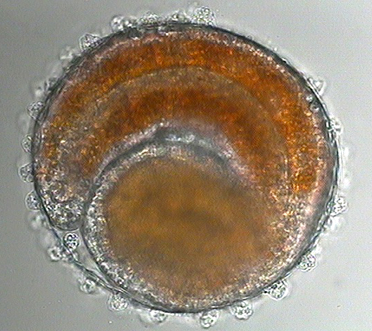
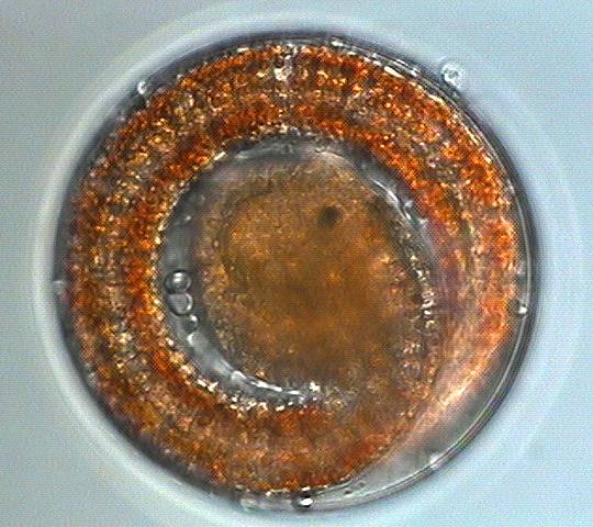
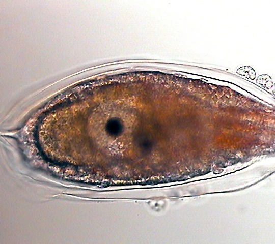
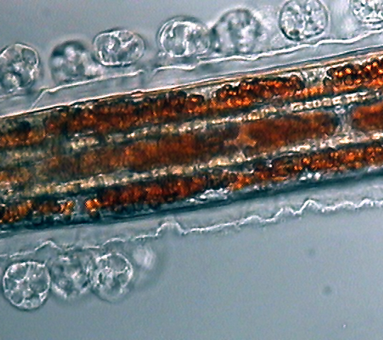
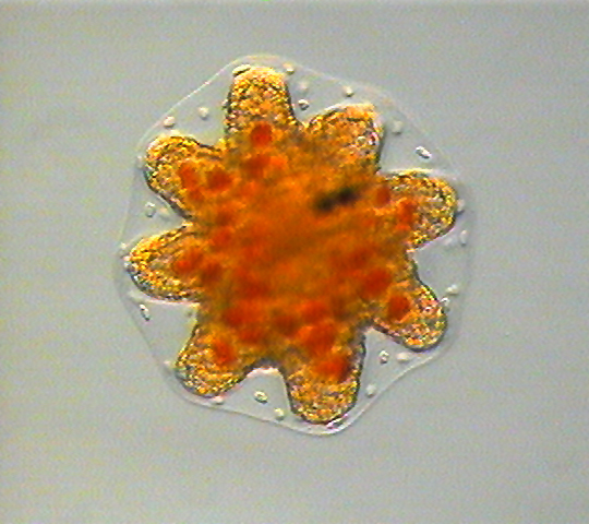
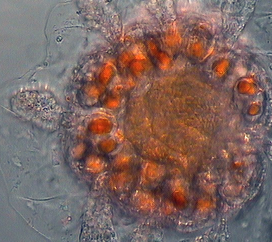
Eggs of the sea squirt Boltenia villosa come out looking normal enough but with a beautiful investment of follicle cells on the outside of the chorion and a poplation of motile test cells inside the chorion. Between fertilization and first cleavage, all the orange pigment granules first sweep to the vegetal pole to form a cap and then, immediately before cleavage, they move again to form a crescent on one side of the egg. The orange cytoplasm is inherited by a subset of cells at each division, and those cells become the larval muscles. At the onset of gastrulation (first and second panel in row 3 below) these cells are lined up in two blacks at the posterior of the blastocoel, and they end up as parallel rows alongside the notochord in the larval tail. The larva loses its tail when it metamophoses (bottom row); the juveniles look a little like the tadpole was dropped on its head from a great height.
|
|
|
|
|
|
|
|
|
|
|
|
|
|
|
|
|
|