Erika Hoyos
In this study I identified a set of genetic interactions that link the EGF receptor/LET-60/Ras and Notch signaling pathways to describe and analyze at a mechanistic level how a group of six vulval precursor cells (VPCs) determine one of three possible cellular fates, primary, secondary and tertiary during C. elegans vulva development. We used a mathematical model based on a system of ordinary differential equations (ODEs) to simulate the intracellular and intercellular interactions of the network, and used the Ingeneue program to visualize the cellular patterns of genetic expression. This model was able to reproduce the gene expression profiles of Notch and MAPKP observed during vulva differentiation in C. elegans. We tested the effect of EGFR internalization, and stabilization by MAPKP in the robustness of the system with respect to random changes in parameter values; finally, we examined how changes in the efficacy by which MAPKP activates Notch degradation may give raise to certain vulva phenotypes, such as vulvaless and multivulva.
|
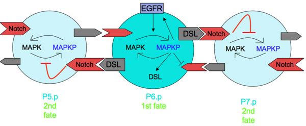
The vulva is patterned from a row of 6 precursor cells that differentiate under the influence of EGF and Notch/Delta signaling. Schematic of interactions is shown here, and the computational model reconstructs these interactions in a computer. |
|
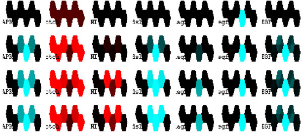
Output of computer simulation for some of the relevant molecules. The simulation shows the time evolution of the different molecules in the system. The columns correspond to (left to right): MAPK activity, Notch, internalized Notch, dsl transcription, lag2 transcription, egf production, and EGF protein levels. The top row corresponds to the initial conditions of the simulation, and successive rows show the state of the system at time intervals separated by 200 minutes. Each hexagon represent a cell in the simulation. Higher levels of activity are shown in brighter colors; different molecules are in different colors. Our model is capable of reproducing the observed pattern (high MAPK activity in the 4th cell from left, lower in its neighbors that also have high NI activity). |